
Super-resolution Light Microscopy
& Nanoscopy Facility (SLN)
The Super-resolution Light Microscopy & Nanoscopy (SLN) Research Facility at ICFO is equipped with front-end microscopy techniques that are able to operate a step beyond the commercial state of the art.
The SLN research team performs continuous R&D in most of the advanced light microscopy techniques and provide access and training to all types of users in the forefront of microscopy for the most demanding biomedical applications. The SLN is open to external collaborations with industry, hospitals, research centers and Universities.
The SLN Facility at ICFO provides:
- Access to external researchers and collaborators to the variety of state-of-the-art microscopy and super resolution techniques.
- Training, through short-term and mid-term, hands-on courses tailored to the needs of specific users with a variety of relevant backgrounds.
- Image analysis and quantification tools customized for the different techniques.
Facility Coordinator
Dr. Pablo Loza- Alvarez
Facility Staff
Eric Calatayud
Dr. Gustavo Castro
Dr. Mónica Marro
Dr. Nicolás Mateos
Research Team in Facility
External Users
Become a user or collaborate with us.
Booking System
Become a user or collaborate with us.
Current Fees
Equipment
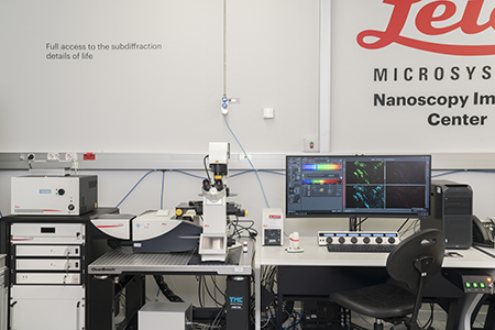
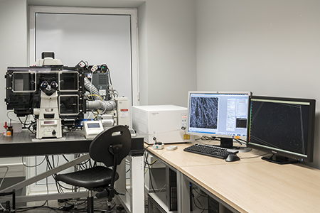
Leica TCS SP8 STED 3X-FALCON
The Leica TCS SP8 STED 3X-FALCON is based on the TCS SP8 confocal microscope. The STED 3X module allows 3D super-resolution capabilities in 3 optical bands due to its 3 depletion lasers. The FALCON (FAst Lifetime CONtrast) is a fluorescence lifetime imaging microscopy (FLIM) platform fully integrated in the confocal microscope that can deliver video-rate FLIM with pixel-by-pixel quantification.

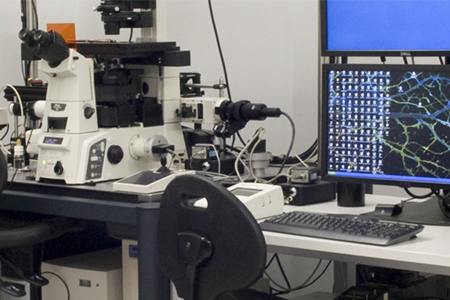
Leica TCS SP5 STED CW
The Leica TCS SP5 STED CW system is based on the Leica TCS SP5 confocal system. It includes the STED CW module. The HyD detectors operate in photon counting mode providing high sensitivity and detection capabilities.
Multimodal Nikon microscope
This is a multimodal workstation built on top of a commercial inverted microscope. This system contains an external scanning head where different femtosecond lasers are coupled.
Renishaw Raman inVia spectrometer system
This is a confocal InVia Renishaw Raman microscope that includes two excitation lasers: 785nm and 532nm.
Multimodal Digital Scanned Light Sheet Microscopy (mDSLM)
The multimodal SPIM/DSLM system is a custom-made microscope designed to explore large microscopic 3D samples such as organoids or small embryos.
N-STORM
Based in a TIRF configuration with 4 colors laser bench. The focusing position stability is given by the Perfect Focus System and it is fully motorized.

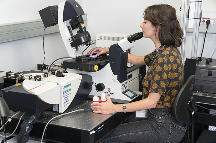
Abberior STED Microscope
Our Abberior STED microscope is based on the INFINITY platform, which is built around an Olympus IX83 microscope. The system is equipped with four pulsed lasers as well as a CW 405nm laser.
Training
We collaborate with industry, hospitals and research centres and we provide training through short and mid-term hands-on courses which can be tailored to accommodate any user needs and backgrounds.
Teaching:
- Master of Multidisciplinary Research in Experimental Sciences.
http://www.bist.eu/master
- Master in Photonics – Optical Experimental techniques in Biology.
http://www.photonicsbcn.eu
- Erasmus Mundus Master Course (EMMC) EUROPHOTONICS in Photonics Engineering, Nanophotonics and Biophotonics.
http://www.europhotonics.org/wordpress/master
- Erasmus Joint Doctorate program.
EUROPHOTONICS EMJD
Partnerships
Research Infrastructures
The SLN forms part of the following research Infrastructures


Collaborations with Industry
ICFO actively collaborates with industry leaders to help implement improvements on the most advanced commercially available microscopy platforms

All SLN Members
Publications Highlights
News Highlights
Funded Projects
Gallery
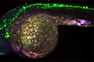
web_ZFish_neurons_stitching_sln-1030×484
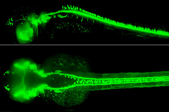
Digital Scanned Laser Light Sheet Microscopy mosaic of a 48 hpf Zebrafish embryo with GFP membrane labelled sensory neurons. Image size 2.8mm x 0.8mm , composition made from 9 stacks of 100 images.
TPEF-ICFO-2
Multicolour two-photon fluorescence imaging is performed using a blue-diode-pumped SESAM-modelocked Ti:Sapphire oscillator generating 5 nJ pulse energy, 82 fs pulse duration, centred at 780 nm, with 92 MHz pulse repetition rate.
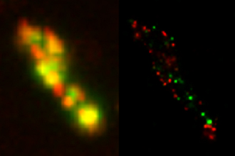
STORM-ICFO-2
HiLo wide field and dSTORM image of Salmonella enterica with RecA protein and CheW labelled with AlexaFluor A647 and Cy3B respectively.
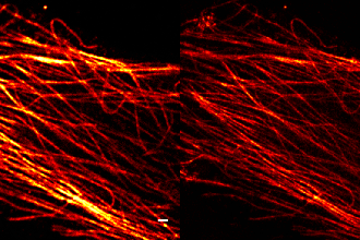
sted_microtubules3
Confocal and STED images of microtubules immunostained with an antibody against alfa-tubulin and a secondary antibody AlexaFluor488. Scale bar 1 µm.
SHG-collagen-TPEF-fibroblasts copia
Multiphoton images of human corneas. In blue, collagen fiber bundles imaged using pSHG. In red, autofluorescence image of fibroblasts using two-photon excitation.
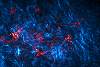
nuclear-pores
Confocal and STED images of nuclear pores immunostained with an antibody against Nup153 and a secondary antibody STAR-488. The comparison shows the improvement in resolution when a CW 592 depletion laser is used to perform STED. Scale bar 1 µm.
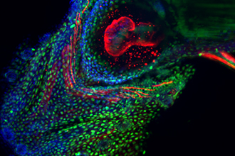
GCAMP_mar-1030×355 copia
Multicolor light-sheet microscopy of a fixed zebrafish larvae expressing GCaMP:GFP on the motor neurons (green). Red and blue channels are autofluorescence (green and red excitation). The image is composed by the stitching of three z-stacks.
20X_1mic_DS_BGDAPI_ventral
Multicolour light-sheet fluorescence microscopy of a fixed zebrafish larva. Nuclei is stained with DAPI (blue), histone with AlexaFluor 488 (green) and cardiac myosin light chain with Alexa Fluor 568 (red).